











Before modern science truly illuminated the process of human reproduction, people held many strange and confident ideas about where life came from. For a long time, new life was believed to arise spontaneously from nonliving matter, as if life itself could simply emerge when the conditions were right. By the middle of the 17th century, this belief began to crack. Natural philosophers were finally able to make out, with the naked eye, the female ovum — the egg cell. A new theory soon appeared: all living beings had already been arranged at the moment of divine creation, one person pre-existing inside another, nested within the woman’s egg like a set of Russian dolls.
This theory came to be known as preformation. It was not merely an idea about biological development; it also happened to flatter the social order of its time. The Portuguese developmental biologist and writer Clara Pinto-Correia observed in The Ovary of Eve that preformation, by placing bloodlines, families and descendants inside one another, could easily become a doctrine that appeared natural while quietly defending hierarchy. It supplied a kind of biological legitimacy to dynastic inheritance and the politics of lineage. And the most influential natural philosophers of the Scientific Revolution were, of course, not speaking from the side of the ruled.
One might imagine that, as microscopes improved and observation became sharper, this Russian-doll vision of life would have been quickly shattered by science. But history rarely advances in so clean a line. Instead, when microscopes finally allowed researchers to see sperm, preformation did not immediately disappear. It merely changed its costume. Where once the future human being had been imagined as hidden inside the egg, some philosophers and early students of reproduction now came to believe that the true blueprint of life was carried by sperm. The egg was reduced to a vessel waiting to receive; sperm, by contrast, was imagined as active, vigorous and filled with the force of life.
In this picture, the head of each sperm contained a tiny, already formed human being. This little figure was called a homunculus. The Dutch mathematician and physicist Nicolaas Hartsoeker, inventor of the screw-barrel microscope, drew the now famous image of this “little man” in 1695, not long after sperm first became visible. Hartsoeker himself admitted that he had not actually seen such a figure inside the sperm head. But that did not prevent him from believing it must be there, just beyond the resolving power of the instruments of his day.
As microscopes grew more powerful, the homunculus was eventually swept into the corner of scientific history, preserved as a cautionary tale about early observation and excessive imagination. Yet the shadow it cast did not vanish completely. Even today, one can still detect its outline in popular accounts of fertilisation: the egg is often portrayed as silent, still and passive; sperm as brave, active and forward-driving. The beginning of life is made to look like a heroic story launched by sperm and merely accepted by the egg.
It is not difficult to understand why the public absorbed this version. The “sperm race” is an easy story to tell: hundreds of millions of sperm charge toward a single egg, and the fastest, strongest and most excellent one wins the prize of fertilisation. But what is unfortunate is that many biologists, physicians and works of science communication have also long relied on this same narrative. In 1991, the American anthropologist Emily Martin famously described this portrayal of egg and sperm as a “scientific fairy tale.” She pointed out that it implied female biological processes were less valuable than their male counterparts, and that women themselves were somehow less worthy than men.
This bias does not live only in metaphor. The ovary is often described as holding a limited supply of eggs, gradually depleted with age, like an inventory running down. The testes, meanwhile, are described as continually producing new sperm, as if the male reproductive system remained permanently abundant. A woman begins puberty with roughly 300,000 egg precursor cells, of which only about 400 will ever mature and be released; this has often been described as “wasteful.” Yet a man may produce more than two trillion sperm over a lifetime, and the same word is rarely applied to that enormous output.
In popular media and scientific writing alike, human fertilisation is commonly presented as a grand long-distance swimming contest. Sperm burst forth, pass through hostile terrain, strain toward the egg, and the quickest, strongest one becomes the victor. The story is vivid, even cinematic. But its problem is that it is not merely a harmless metaphor. It repackages reproduction as a story of male competition, male triumph and male initiative, while obscuring the complexity of the real biological process.
If this were only an outdated fantasy inherited from a sexist past, that would already be bad enough. More seriously, when a misleading narrative is mistaken for scientific fact, it affects how we understand fertility, how we diagnose infertility, how we design assisted reproductive technologies, and where we direct attention — toward men, toward women, or toward the interaction between the two. To see how this myth took shape, we must return to the long history of how science came to understand human reproduction.
Our knowledge of sex cells and conception is, in fact, quite recent. The egg is the largest cell in the human body, barely visible to the naked eye, about the size of the period at the end of a sentence. Sperm, by contrast, is one of the smallest human cells and cannot be seen without a microscope. It was not until 1677 that the Dutch amateur scientist Antonie van Leeuwenhoek first observed human sperm under magnification. Around the same time, people began to realise that the human ovary produced eggs, but actual observations of human and other mammalian eggs were not reported until the work of the German biologist Karl Ernst von Baer in 1827.
Even after sperm had been discovered, its role was not immediately understood. Nearly a century after van Leeuwenhoek’s observation, someone finally demonstrated that sperm was necessary for fertilisation. In the 1760s, the Italian priest and natural scientist Lazzaro Spallanzani performed experiments with frogs. He fitted male frogs with tight little trousers made of taffeta, preventing sperm from entering the water, and found that the eggs did not develop into tadpoles. The experiment may sound comical, but it was a major step in the history of reproductive science.
Before that, many people believed sperm were merely tiny parasites living in semen. Even van Leeuwenhoek himself accepted a similar view for a time. It was not until 1876 that the German zoologist Oscar Hertwig observed and demonstrated the fusion of sperm and egg in sea urchins, allowing fertilisation to be understood gradually as the union of two sex cells.
Better microscopes eventually revealed a startling fact: a typical human ejaculate, with a volume of only about half a teaspoon, may contain roughly 250 million sperm. This raised a deceptively simple question, one that has never been answered as fully as it should be: why so many?
Studies show that when the sperm count in a single ejaculate falls below 100 million, pregnancy rates tend to decline. In other words, a substantial proportion of the sperm in an average ejaculate does seem to be connected to normal fertility. For a long time, the most popular explanation was “sperm competition.” According to this view, sperm exist in such huge numbers because they must compete with one another for access to the egg; the more sperm there are, the greater the chance of winning the fertilisation prize, rather like buying more tickets in a lottery. If a woman might mate with more than one man, then sperm from different men could also compete. Natural selection, in this story, drives men to produce more sperm in an arms race for the chance to fertilise.
Sperm competition certainly exists in the animal kingdom, and sometimes in striking form. Chimpanzees are a classic example. As one of our closest living relatives, chimpanzees live in groups that include multiple adult males, and females often mate with several males. In such an environment, male competition does not occur only through physical confrontation; it also takes place inside the female body, at the level of sperm. Chimpanzees have relatively enormous testes, rapid sperm production, high sperm counts, and large sperm midpieces packed with mitochondria that supply energy for movement. Their sperm-carrying ducts are muscular, their seminal vesicles and prostate glands are well developed, and their semen can coagulate to form a plug that temporarily blocks access by the sperm of other males. Higher white blood cell counts also help counter the risks of sexually transmitted pathogens that come with frequent mating.
Such examples are compelling, which is why it has been so tempting to apply the same model to humans. The problem is that humans do not appear to be strongly adapted for intense sperm competition in the way chimpanzees are. Despite the lurid claims that circulate in popular culture about male competition, sexual strategy and sperm wars, the evidence does not support the conclusion that human males evolved primarily under strong sperm-competition pressure.
Compared with chimpanzees, humans are closer to primates that live in groups with a single breeding male. In such species, direct sperm competition is limited and testes are relatively small. Human testes are about the size of walnuts, roughly a third the size of chimpanzee testes, which are closer to the size of chicken eggs. More importantly, chimpanzee semen contains a relatively low proportion of physically abnormal sperm, whereas human semen contains a large number of structurally defective sperm. If humans were truly under strong sperm-competition pressure, we might expect stricter quality control. Instead, we see the opposite. Human semen contains many “duds,” suggesting that direct sperm competition is not the best explanation for human sperm numbers.
The Journey of Sperm Is More Like a Layered Obstacle Course Than a Swimming Race
If humans are not obviously adapted for fierce sperm competition, how else might we explain the existence of so many sperm? One less familiar but illuminating possibility comes from the biologist Jack Cohen at the University of Birmingham. More than four decades ago, in a few papers that have rarely received the attention they deserve, Cohen proposed that sperm numbers might be connected to chromosomal recombination during sperm production.
In meiosis, the special kind of cell division that produces sperm and eggs, paired chromosomes exchange pieces of genetic material through crossing over. This process increases the diversity of genetic combinations, and such diversity is the raw material on which natural selection can act. Cohen noticed that, across species, sperm numbers appeared to be associated with the number of chromosomal crossovers occurring during sperm production. In other words, producing many sperm may not be only about competition. It may also be a way of representing many different genetic combinations.
One can imagine sperm production as a kind of lottery system. The more available numbers there are — that is, the more possible genetic combinations — the more tickets must be printed, meaning the more sperm must be produced. This comparison is less dramatic than the familiar sperm race, but it may be closer to the genetic logic of reproduction.
Many other findings also undermine the classic story of sperm swimming heroically all the way to the egg. In most mammals, sperm do not travel through the entire female reproductive tract solely by their own power. For much of the distance, they are transported passively by contractions, waves and fluid movements in the uterus and oviducts. Sperm do move, of course, but they are not lone athletes swimming from start to finish.
A simple comparison makes the point. Sperm in smaller mammals are often longer, on average, than sperm in larger mammals; mouse sperm are longer than whale sperm. Yet the larger the animal, the longer the female reproductive tract. If sperm movement were understood entirely as independent swimming, then whale sperm would face a journey far beyond that of mouse sperm — an implausible scenario. In many large mammals, sperm simply could not reach the fertilisation site without the assistance of the female tract’s transport mechanisms.
Humans are no exception. Increasing evidence indicates that human sperm are passively transported over considerable distances as they pass through the uterus and into the oviducts. The idea of the Olympic-champion sperm looks, in this light, not only simplistic but excessively heroic in a masculine key.
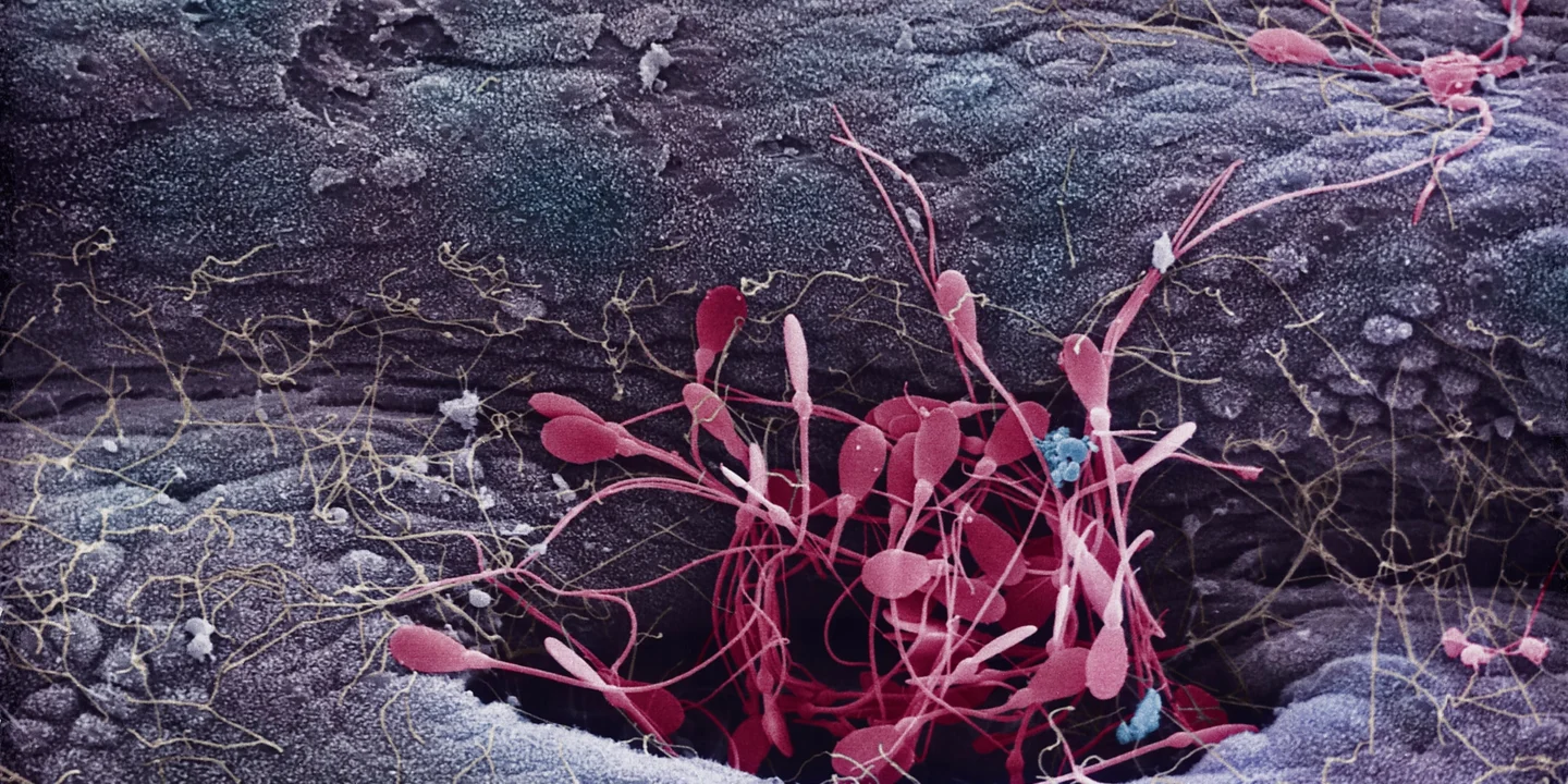
In fact, from an ejaculate containing around 250 million sperm, only a few hundred will eventually reach the upper oviduct and come near the egg. The process is not a standard swimming race but a brutal obstacle course. Sperm numbers are cut down layer by layer within the female reproductive tract, until fewer than one in a million of the original sperm are present near the egg at the time of fertilisation.
Many sperm never even enter the cervix. The vagina is acidic and hostile to sperm, which cannot survive there for long. Those that escape the vagina and reach the cervix must still face the filtering action of cervical mucus. Structurally abnormal sperm are more likely to be trapped. At the same time, large numbers of sperm enter side channels in the cervix, known as crypts, where they may be stored for several days. Only a minority of sperm travel directly through the uterine cavity and continue upward.
Even after entering the oviduct, the filtering is not over. Sperm may temporarily bind to the inner surface of the oviduct, and only some are later released to approach the egg. The female reproductive tract, then, is not an empty tube passively awaiting sperm. It is a complex system of selection, storage, transport and restriction. It helps sperm move forward, but it also prevents too many sperm — or defective sperm — from approaching the egg.
This is why the story of “the best sperm wins” is so misleading. It makes us overlook an equally important fact: more sperm is not always better. If too many sperm gather around the egg, the risk of polyspermy increases. Polyspermy occurs when more than one sperm enters the same egg. Its consequences are usually severe.
In humans, polyspermy occasionally occurs, especially when the father has an unusually high sperm count. The most common result is that two sperm fertilise one egg, producing an embryo whose cells contain 69 chromosomes instead of the normal 46. This condition is almost always fatal and usually results in miscarriage. Even in the rare cases in which development continues until birth, survival is typically brief.
From an evolutionary perspective, then, the many barriers in the female reproductive tract are not accidental inconveniences. They are important mechanisms that help prevent polyspermy and maintain normal fertilisation. Natural selection does not merely encourage males to produce more sperm; it also shapes female systems that limit sperm numbers. The beginning of life is not a one-sided male charge, but a delicate balance between structures and processes that evolved in both sexes.
This understanding matters for assisted reproductive technologies as well. Early artificial insemination often involved placing semen in the vagina. Later, intrauterine insemination, or IUI, became more common: processed sperm are placed directly into the uterus. This bypasses part of the natural filtering that normally occurs in the cervix, including the trapping of abnormal sperm by cervical mucus. For that reason, sperm numbers in IUI must be handled carefully. Clinical data suggest that placing around 20 million sperm in the uterus is enough to achieve a routine pregnancy rate — less than one-tenth of the sperm count in an average ejaculate.
In in vitro fertilisation, or IVF, the issue becomes even more obvious. IVF exposes eggs directly to sperm in a laboratory dish, bypassing the entire series of natural filters from the vagina to the cervix, uterus and oviduct. In the early development of IVF, large numbers of sperm were often used in the hope of increasing fertilisation success. The logic seemed reasonable: more sperm should mean a greater chance of success. But the reality is more complicated. Excessively high sperm numbers can actually reduce success rates and may increase the risk of polyspermy. Studies have indicated that fertilisation works better when around 25,000 sperm are placed near an egg.
This reminds us that a bad metaphor is not merely a linguistic problem. It can shape technical choices, influence how physicians and researchers understand risk, and determine where attention is directed.
Human Fertilisation Is More Like a Vast Drawing of Lots: Among Healthy Sperm, Success Often Depends on Chance
Once the danger of polyspermy is taken seriously, our understanding of the evolution of sperm numbers changes. Traditional discussions of sperm competition usually focus on maximising sperm count, as if more sperm always gives males an advantage. But biology rarely offers advantages in only one direction. Every increase comes with a constraint; every form of competition carries a cost. Males may evolve higher sperm production in some environments, but the female reproductive tract may also evolve mechanisms to control sperm numbers and protect the egg.
In promiscuously mating primates such as chimpanzees, longer female oviducts may partly offset higher male sperm production. Males produce more sperm; female reproductive anatomy, in turn, limits how many of those sperm can finally reach the egg. This means the female is not a passive arena for sperm competition. She is an active participant in an evolutionary negotiation.
The idea that “the best sperm wins” remains stubbornly attractive because it satisfies our preference for competition stories. It makes fertilisation look like sport: there is a starting line, a finish line, a field of rivals and a champion. But from the standpoint of cell biology, this story is difficult to sustain. The DNA inside a sperm head is tightly compressed, almost crystalline. How could an external system assess whether the genes inside one sperm are better or worse than those inside another? How could an egg, or the female reproductive tract, detect the genetic quality of a sperm before fertilisation?
Experiments do not support many popular speculations. Some have imagined, for example, that the female reproductive system might favour sperm carrying an X chromosome or a Y chromosome, thereby influencing the sex of the offspring. But experiments in mice have not provided clear evidence for such selection. Among sperm that are structurally normal and functionally healthy, the one that fertilises the egg may not be the “best” in any meaningful sense. It may simply be the one that happens to be in the right place at the right time.
Human fertilisation, then, may be better understood as a vast drawing of lots with 250 million tickets. Large sperm numbers represent a large set of possible genetic combinations. The female reproductive tract filters out obvious defects, limits total numbers, stores and releases some sperm, and allows only a tiny fraction to approach the egg. Among the healthy sperm that remain, there may be no sophisticated ranking system. Which one succeeds may depend, to a large degree, on chance.
The large proportion of structurally abnormal sperm in human semen has also long puzzled researchers. Some sperm have two tails; some have tiny heads; some are visibly malformed. One once-famous hypothesis proposed that these abnormal sperm were not useless after all, but rather “kamikaze sperm.” Their job, according to this theory, was not to fertilise but to block, interfere with, or even kill the sperm of other men. The story was dramatic and fit neatly into a male-war narrative. But later evidence has not supported it, and the idea has largely been discredited.
Another long-neglected fact is that sperm do not necessarily rush straight to the egg after entering the female reproductive tract. Many may be stored, remaining in place for days before being gradually released. For a long time, it was widely believed that human sperm could survive inside the female tract for only two days. But from the mid-1970s onward, accumulating evidence showed that sperm can remain intact for at least five days. Today, extended sperm survival is widely accepted, and in some cases it may last ten days or even longer.
This has important implications for so-called “safe period” or “natural” methods of birth control. Many such methods depend on the assumption that sperm survival is short and that avoiding intercourse close to ovulation is enough to prevent conception. But if sperm can be stored and remain viable in the female reproductive tract for much longer, then the fertile window is not as narrow as once imagined. The risk of conception correspondingly increases.
Cervical mucus plays a central role here. People often talk about changes in the state of cervical mucus, especially in natural birth-control methods. Near ovulation, the mucus becomes thinner, wetter and more slippery, and this is treated as a sign of fertility. Yet far less attention has been paid to how cervical mucus participates in sperm storage.
In fact, sperm can be stored in cervical crypts, the very structures from which mucus is secreted. Unfortunately, direct research on this process is extremely limited. In 1980, the gynaecologist Vaclav Insler and colleagues at Tel Aviv University published an important study. Twenty-five women, scheduled to undergo hysterectomy the following day, volunteered to be artificially inseminated. After surgery, the researchers examined serial sections of cervical tissue under the microscope to observe where sperm had gone.
The results showed that within two hours of insemination, sperm had colonised the entire length of the cervix. The cervical crypts varied greatly in size, and sperm were concentrated mainly in the larger ones. In some women, as many as 200,000 sperm were stored in the cervical crypts. The researchers also reported that live sperm had been found in cervical mucus as late as the ninth day after insemination.
This was a striking finding. It suggested that the cervix is not merely a passageway for sperm but may function as a sperm reservoir. Sperm remain there, stay viable, and are gradually released to continue toward the oviduct. Strangely, although this study has been widely cited, it has rarely been absorbed into the mainstream story of fertilisation, and systematic follow-up studies have been scarce. Sometimes science does not lack evidence; rather, some evidence fails to fit the story we have grown used to telling, and so it remains on the margins.
Mutations Accumulate Faster in Sperm: Male Fertility Is Not Untouched by Time
The British physiologist Sir Robert Edwards was a central figure in the development of IVF and later received the 2010 Nobel Prize for that work. In his 1980 textbook Conception in the Human Female, a work of more than 1,000 pages, he discussed human conception in extraordinary detail. Yet sperm storage in cervical crypts received only a brief mention. Since then, many other authors have referred to the phenomenon only in passing.
But sperm storage and gradual release are crucial for understanding human reproduction. They affect not only how we think about ovulation, the fertile window and contraceptive risk, but also how we approach infertility treatment. If sperm can be stored in the cervix and remain viable, then the female reproductive tract is not merely a filter. It is also a timing system. It regulates when sperm move forward, how many move forward, and which ones move forward. This complexity far exceeds the simple image of sperm swimming heroically toward the egg.
There is another persistent misunderstanding: the myth of male fertility as essentially unaffected by age. Popular culture often assumes that female fertility declines sharply with age and ends at menopause, while men can continue reproducing into old age with little biological consequence. This contrast is incomplete and inaccurate.
A large body of evidence shows that sperm count and sperm quality decline as men age. More importantly, recent research has indicated that mutations accumulate in sperm at roughly four times the rate seen in eggs. One reason is that sperm production requires continuous cell division, and the more often cells divide, the more opportunities there are for copying errors and mutations to build up. Semen from older men, therefore, is not risk-free. It may carry a higher mutational burden and may affect the health of offspring.
In modern industrialised societies, the increasing age at which women have their first child has generated extensive discussion. People repeatedly talk about the risks of older motherhood, declining egg quality and “fertility preservation.” One frequently proposed solution is for young women to freeze their eggs for later use. But egg freezing is invasive, expensive and physically demanding, requiring hormonal stimulation, egg retrieval and long-term storage.
By contrast, the reproductive problems associated with ageing men have received far less attention. If mutations accumulate faster in sperm, then one simpler, cheaper and far less invasive strategy would be for young men to store semen samples for future use. Sperm freezing is already a mature technology and is much easier than egg retrieval. Yet in public discussion, this option is rarely placed on the same level of importance as egg freezing.
This is not because male age is irrelevant. It is because our culture has long been accustomed to placing reproductive risk on women. Women are treated as the ones who must manage the biological clock, while men are imagined as providers of fertility whose reproductive capacity simply continues. This imagination is neither fair nor scientific. It places excessive pressure on women and underestimates risks connected to men.
Looking back, Hartsoeker’s little man inside the sperm may seem very distant. No one today seriously believes that a complete tiny human being curls inside the sperm head, waiting to enter the egg and grow into a person. The image survives more as a relic of scientific history, a curious anecdote about early microscopes, imagination and misplaced confidence.
But the real legacy of the homunculus does not lie in whether the theory itself was overturned. It lies in the way of thinking it represented. We still easily imagine the male cell as the active party and the female cell as the container; sperm as heroes and the egg as the prize; fertilisation as competition rather than complex interaction.
Science does not ask questions in a vacuum. It always unfolds within a cultural language, and it is always vulnerable to the influence of social imagination. Research on human reproduction is especially susceptible, because it is naturally entangled with sex, family, lineage, bodies and power. When we repeatedly tell the story of the macho sperm, we misunderstand not only sperm but also eggs, the female reproductive tract, the risks of male ageing, and what truly happens at the beginning of life.
A more reliable reproductive science must move beyond metaphors that are vivid but biased. It must recognise that the egg is not a passive prize, the female reproductive tract is not an empty corridor, and sperm are not a crowd of swimmers racing toward a podium. Fertilisation is not a contest of male victory. It is a precise, fragile biological process full of selection, restriction, timing and chance.
Reducing gender bias is not merely a matter of fairer language. It can help us ask better scientific questions, design more rational treatments, and assign reproductive responsibility more honestly and equally to men and women alike. The myth of the macho sperm has lasted far too long. Like the little man once imagined inside the sperm head, it belongs, at last, in history.